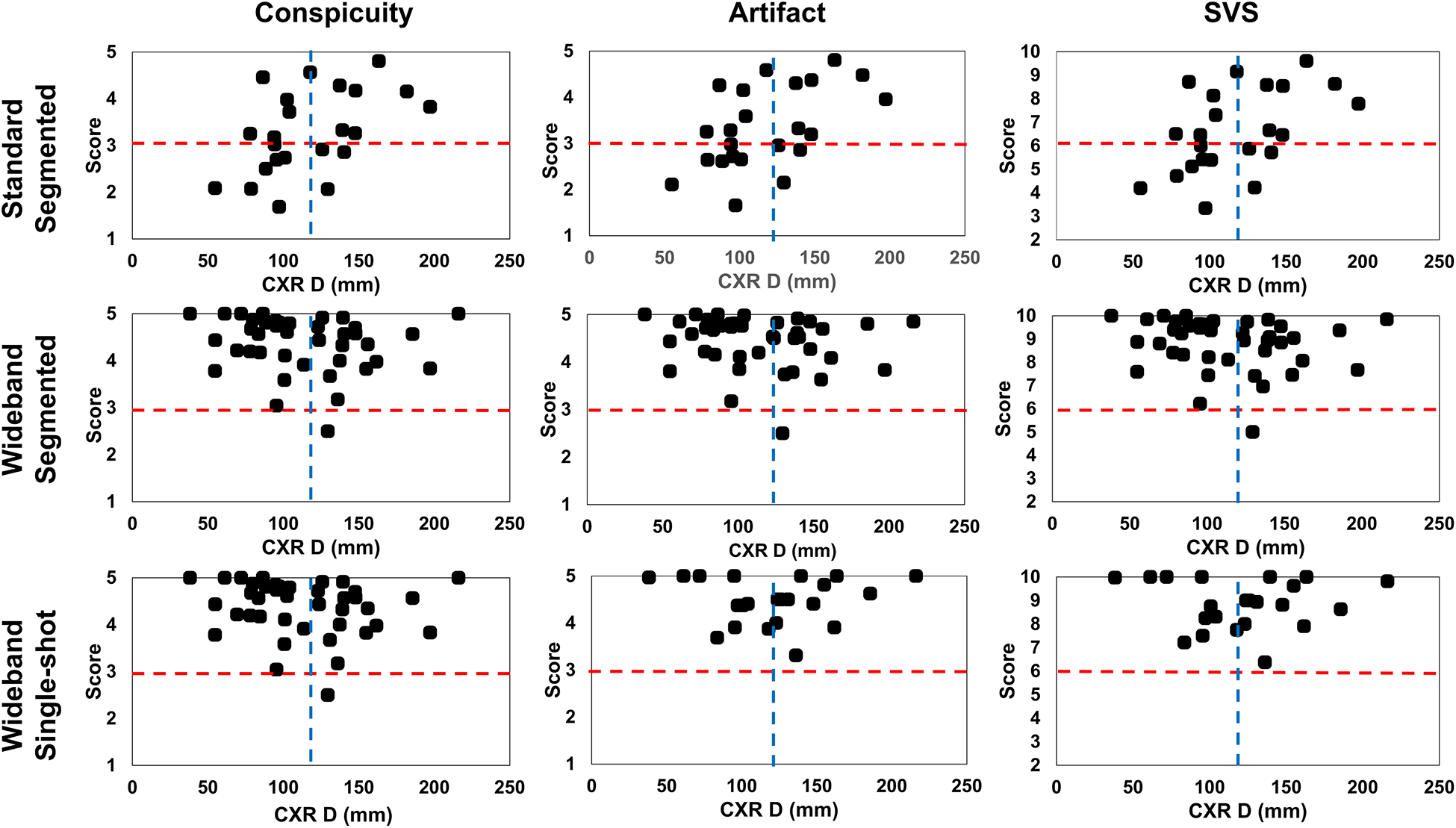
Figure 5:

Segmental conspicuity, artifact, and SVS in 16 myocardial segments across pulse sequences using a Kruskal-Wallis test. As shown, segmental scores varied more for standard LGE than wideband segmented LGE and wideband single-shot LGE. These plots exclude data from four patients with S-ICD, which is implanted in a different location and transvenous devices. SVS: summed visual score; S: subcutaneous.
