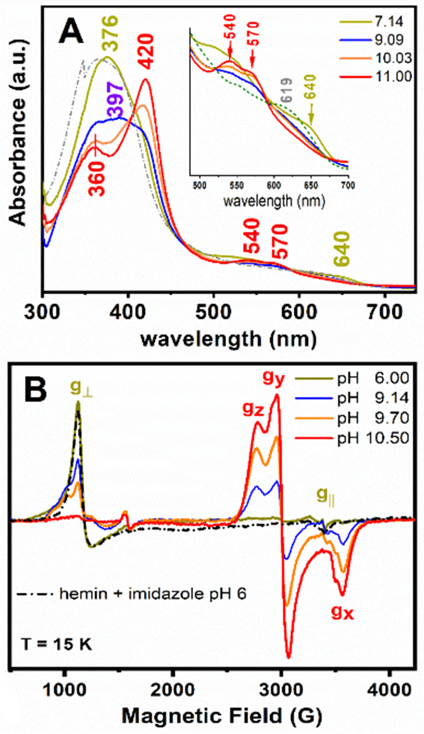
Figure 3.

UV-Vis electronic absorption (Panel A) and 9-GHz EPR (Panel B) spectra of the GRW-L16CL30H mini-heme protein (15 μM and 300 μM heme concentration, respectively; one heme per peptide dimer). The absorption spectrum of hemin in buffer solution (pH 7, dash-dotted trace) and the EPR spectrum of hemin in imidazole buffer (pH 6, dash-dotted trace) are also shown. The pH-induced spectral changes were fully reversible. Freezing and thawing the EPR sample did not affect the ligation and the reversibility in heme ligation. EPR experimental conditions as in Fig. S6.
