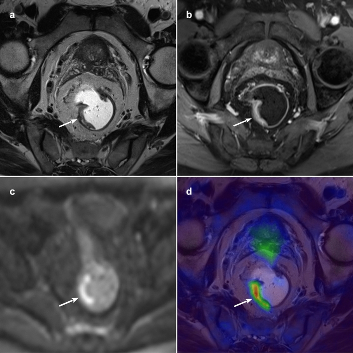
Fig. 2.
PET/MR images of a histologically-proven T2 rectal cancer after pCRT. a T2-weighted paraxial image showing an irregular thickening of the right rectal wall (arrow), with no signs of extramural invasion; b paraxial contrast-enhanced volumetric interpolated breath-hold examination (VIBE) with irregular enhancement of the same lesion (arrow); c axial b1000 diffusion-weighted imaging (DWI) showing signal restriction of the mass (arrow); d paraxial PET/MR fused image with hypermetabolism of the rectal tumour (arrow)

