Figure 2.
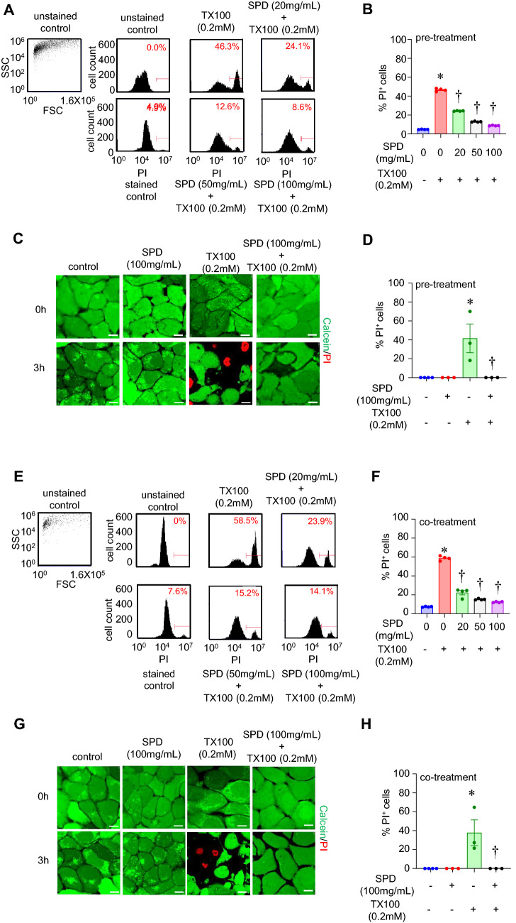
Pre- and co-treatment of human keratinocytes with SPD protected against TX100-induced cell death. (A–D) Human keratinocytes (HaCaT) were pretreated with SPD (100 mg/ml) for 24 h and then exposed to TX100 (0.2 mM) for 3 h in the continued presence of SPD (100 mg/ml). (A,B) Cells were stained with PI for flow cytometry. Flow cytometry histograms and quantification of %PI-positive cells. Data represent mean ± SEM (n = 4). *p < 0.05 compared to control (TX100-untreated). †p < 0.05 compared to TX100-treated group. (C,D) Cells were stained with Calcein and PI for confocal microscopy. Representative images and quantification of %PI-positive cells are shown. Data represent mean ± SEM (n = 3-4). *p < 0.05 compared to control (TX100-untreated). †p < 0.05 compared to TX100-treated group. (E–H) Human keratinocytes (HaCaT) were co-treated with SPD (100 mg/ml) and TX100 (0.2 mM) for 3 h. (E,F) Cells were stained with PI for flow cytometry. Flow cytometry histograms and quantification of %PI-positive cells. Data represent mean ± SEM (n = 4). *p < 0.05 compared to control (TX100-untreated). †p < 0.05 compared to TX100-treated group. (G, H) Cells were stained with Calcein and PI for confocal microscopy. Representative images and quantification of %PI-positive cells are shown. Scale, 10 µm. Data represent mean ± SEM (n = 3-4). *p < 0.05 compared to control (TX100-untreated). †p < 0.05 compared to TX100-treated group.

