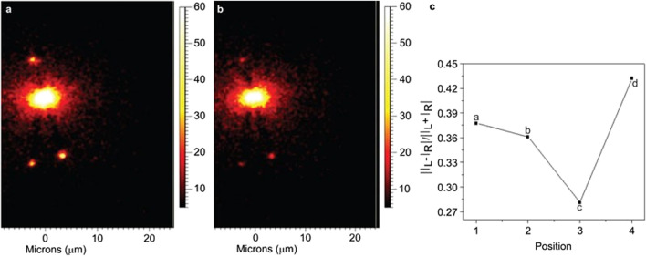
FIGURE 7.
Imaging of the Raman peak of FGGO at 1593 cm−1, excited by circular polarization, and the fitted CIDs at four different points. (A) Results for left circular polarization. (B) Results for right circular polarization. (C) The fitted CIDs at four points of the nanostructure. CID, circular intensity difference; FGGO, fmoc-glycyl-glycine-OH. The figure has been adapted with permission from Sun et al., (2013), 2013 CIOMP.

