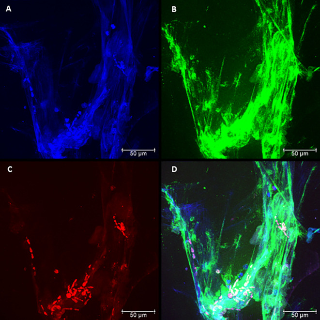
Figure 2.
Confocal microscopy of neutrophils challenged with P. brasiliensis - Pb265 (50:1 ratio), showing the pattern of neutrophil extracellular traps (NETs) release. Cocultures were stained with DAPI (A), labeled with anti-elastase antibody followed by FITC-conjugated secondary antibody (B), and anti-histone H1 secondary antibody followed by Texas Red (C). In the last frame, the overlapping images showing the three components of NETs (D). (Bar size 50 μm). The images are reprentative of 10 individuals tested.

