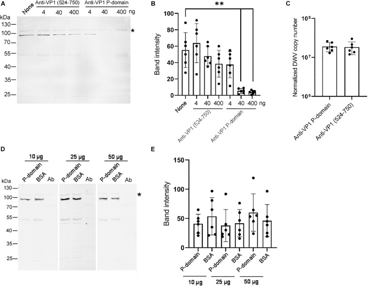
FIGURE 3.
Essential role of the VP1 P-domain in DWV infection. (A) Effect of preincubation of DWV with 4, 40, or 400 ng of either anti-VP1 (524–750) or anti-VP1 P-domain antibody before infection on RdRP synthesis. DWV with no antibody preincubation (none) was used as a control. (B) Band intensity of the RdRP precursor was compared between the seven groups with the one-tailed Dunnett test. P-values between none and 40 or 400 ng of anti-VP1 P-domain antibody are <8.3 × 10⁻⁶ and <5.5 × 10⁻⁶, respectively (**). Mean values ± SD (error bars) are shown (n = 6). (C) Entry of DWV pre-incubated with 40 ng of either anti-VP1 (524–750) or anti-VP1 P-domain antibody to the pupal head tissues was compared (normalized copy number of DWV inside the infected cells). The difference between the two groups was not statistically significant. Mean values ± SD (error bars) are shown (n = 6). (D) Effect of preincubating pupal head tissues with 10, 25, or 50 μg of purified P-domain protein before DWV infection on RdRP synthesis. BSA was used as control. Lysate of abdomen from the same pupa (Ab) was also analyzed to confirm the lack of replication of endogenous DWV. (E) Band intensity of RdRP precursor was compared between P-domain and BSA with the different amount of protein. The difference between the two groups was not statistically significant. Mean values ± SD (error bars) are shown (n = 6). *This represents statistical significance.

