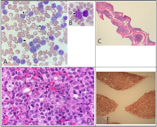
Figure 1.
(A) Peripheral smear: atypical lymphocytosis (black arrows) with deeply folded nuclei and condensed chromatin, scant agranular blue cytoplasm. (B) Hallmark cells: hallmark cells (black arrow) with ‘flower like’ nuclei (not from our patient18). (C) Bone marrow biopsy: low power: hypercellular marrow with decreased maturing lineage haematopoiesis. (D) Bone marrow biopsy: high power: hypercellular (>90%) due to atypical, diffuse, sheet-like infiltrate of lymphoid cells, like those seen in peripheral blood. (E) CD30 staining: strong CD30 positivity in bone marrow.

