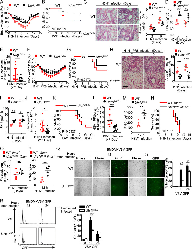
Figure 2.
Uhrf1 deficiency potentiates the antiviral immune response. WT and Uhrf1MKO mice (6–8 wk) were i.n. infected with a sublethal dose (0.1 HA) of H5N1 influenza virus. (A and B) The body weight loss (A) and survival rate (B) were measured for 14 d (n = 20). (C) H&E staining of lung tissue sections on days 2 and 5 after infection. Inflammation scores are presented as a bar graph (n = 5). Scale bar, 200 µm. (D) ELISA for IFN-β in the sera of WT and Uhrf1MKO mice infected with H5N1 influenza virus on days 2 and 5 (n = 3). (E) The viral titers in the lung were quantified on day 2 using the TCID50 assay (n = 10). (F–J) WT and Uhrf1MKO mice (6–8 wk) were i.n. infected with a sublethal dose (0.1 HA) of PR8. The body weight loss (F) and survival rate (G) were measured for 14 d (n = 12). H&E staining of lung tissue sections (H; n = 5) and IFN-β in serum (I; n = 4) was measured on days 2 and 5 after infection. Scale bars, 200 µm. (J) The viral titers in the lung were quantified on day 2 using the TCID50 assay (n = 10). (K–M) Survival rate (K; n = 12), viral titer (L; n = 8), and IFN-β in serum (M; n = 4) of WT and Uhrf1MKO mice intravenously injected with HSV-1 (3 × 10⁶ PFU per mouse). (N–P) WT and Uhrf1MKO mice bred to the Ifnar1−/− background were i.n. infected with a sublethal dose (0.1 HA) of PR8. The survival rate (N; n = 16), viral titer (O; n = 5), and IFN-β in serum (P; n = 4) were monitored for 14 d. WT or Uhrf1-deficient BMDMs were infected with GFP-expressing VSV (VSV-GFP) at an MOI of 0.1 for 24 h. (Q) The data are presented as a representative picture, showing the infected (GFP+) and total (bright-field) cells. Scale bar, 1,000 µm. (R) Summary graph of flow cytometric quantification of the infected cells. All the data are representative of at least three independent experiments. The data are presented as means ± SEMs. The significance of differences was determined by a t test. *, P < 0.05; **, P < 0.01; ***, P < 0.005.

