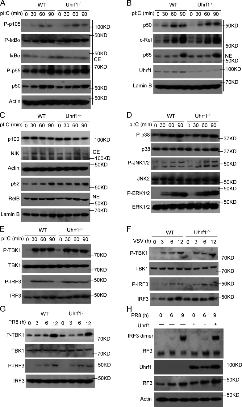
Figure 4.
Uhrf1 does not affect canonical antiviral signal transduction. (A–E) The indicated proteins in cytoplasmic (CE) and nuclear (NE) extracts (A–C) or whole-cell lysates (D and E) of WT and Uhrf1-deficient BMDMs were measured by IB analysis. (F and G) Specific virus-induced phosphorylation of TBK1 and IRF3 in whole-cell lysates was measured by IB analysis. (H) IB analysis of monomeric and dimeric IRF3 (top blot), total IRF3, Uhrf1, and actin (bottom) in HEK293T cells transfected with empty vector or an expression plasmid for Uhrf1 and then infected with PR8 for various times. IB analysis of monomeric and dimeric IRF3 (top blot), total IRF3, Uhrf1, and actin (bottom) in HEK293T cells transfected with empty vector or an expression plasmid for Uhrf1 and then infected with PR8 for various times. All the data are representative of at least three independent experiments.

