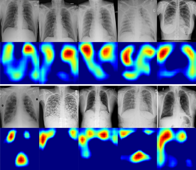
Fig. 1.
Saliency maps for the correctly classified COVID-19 (top two rows) and Non-COVID-19 (bottom two rows) images by the proposed model. Notice that for images that are classified as COVID-19, our model highlights the areas within the lungs, whereas for Non-COVID-19 images, the most important regions are around the lungs

