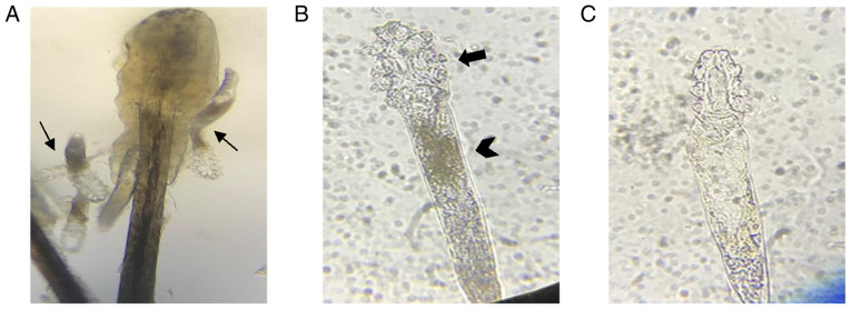
Figure 2.
Microscopic examination of Demodex mites. (A) Several Demodex mites were detected under microscopic examination of the epilated lash (magnification, x20; thin arrows). (B and C) The one of the Demodex mite from (A) was photographed for live and dead status under a microscope (magnification, x40). (B) The Demodex mite was determined to be ‘alive’ if marked movements of the legs (thick arrow) and body (thick arrowhead) was present. (C) By contrast, the Demodex mite was determined to be ‘dead’ when movements of the leg ceased and the body became transparent.

