Figure 2.
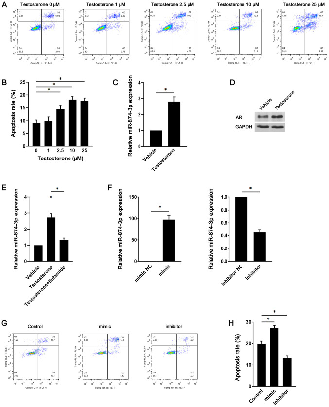
miR-874-3p serves a role in testosterone-induced GC apoptosis. Following treatment with different doses of testosterone for 24 h, mouse GC apoptosis was (A) determined via flow cytometry and (B) quantified. (C) Following treatment with vehicle or 10 µM testosterone for 24 h, miR-874-3p expression levels were detected via RT-qPCR. (D) AR protein expression levels were detected via western blotting. (E) Following treatment with 10 µM testosterone in the presence or absence of 1 µM flutamide for 24 h, miR-874-3p expression levels in mouse GCs were detected via RT-qPCR. (F) Transduction efficiency of miR-874-3p mimic and miR-874-3p inhibitor in mouse GCs. Following transduction with lentiviral vectors expressing miR-874-3p mimic or miR-874-3p inhibitor for 1 day, then treatment with 10 µM testosterone for 24 h, mouse GC apoptosis was (G) determined via flow cytometry and (H) quantified. *P<0.05. miR, microRNA; GC, granulosa cell; RT-qPCR, reverse transcription-quantitative PCR; AR, androgen receptor; NC, negative control.

