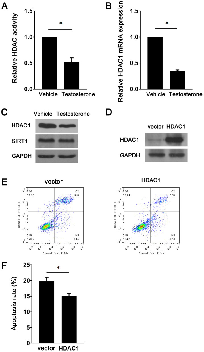
Figure 3.
HDAC1 inhibits testosterone-induced GC apoptosis. Following treatment with 10 µM testosterone for 24 h, (A) HDAC activity, (B) HDAC1 mRNA expression levels, and (C) HDAC1 and SIRT1 protein expression levels in GCs were assessed. (D) At 48 h post-transduction with the HDAC1 overexpression vector or the control empty vector, HDAC1 protein expression levels in GCs were detected via western blotting. Following transduction with the HDAC1 overexpression vector or control empty vector and treatment with 10 µM testosterone for 24 h, mouse GC apoptosis was (E) determined via flow cytometry and (F) quantified. *P<0.05. HDAC, histone deacetylase; GC, granulosa cell; SIRT1, sirtuin 1.

