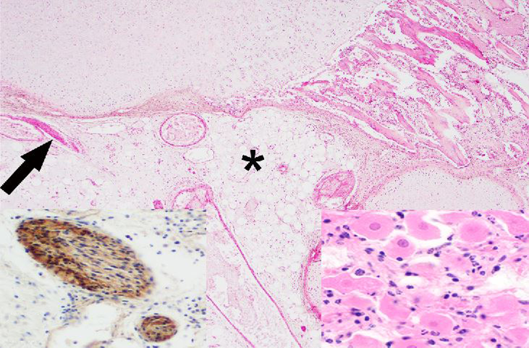
Figure 19. Teratoma, left ovary, rhesus macaque.

The core of the neoplasm is composed of disorganized adipose tissue (*), blood vessels, few nerves (arrow), bone, cartilage (top of image) and a small cluster of neurons (right inset). HE. Left inset: Diffuse cytoplasmic immunolabeling of nerve fibers for S-100.
