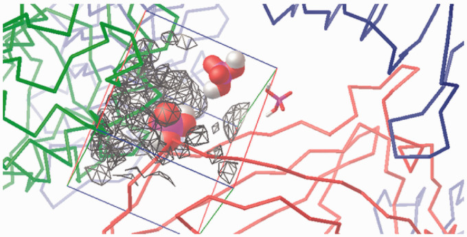
Figure 12.
Visualisation of the –1 charge attraction fields from residues in human wedge calculated by AD4 (for a 40 Å×40 Å×40 Å box). TLR4 (green ribbons, right) counter TLR4* (red ribbons, left) and MD-2 (light blue ribbons, bottom) contour the approximately triangular cavity. The three sites are marked with a H2PO4– mono-anion: Pan (VDW volume, left), PagPan (sticks, right) and Pag (half space-filled, top). In the presence of TLR4*, there is a strong repulsive force at Pan. Hence, a complete human wedge (Δ) is likely to exist only for agonistic LPS-like ligands occupying Pag and PagPan with their dephosphorylated diglucosamine backbone. The triangular shape of the wedge is formed by the three phosphate interaction sites: (a) Pag, on the top right corner of the box; (b) PagPan way down, buried in the cleft to its right, almost centre; and (c) Pan, leftmost phosphate model, which lies halfway on the z-axis (third dimension in the flat picture) between PagPan at the bottom and Pag at the top.

