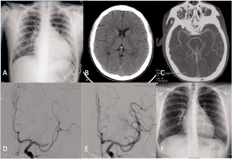
Figure 2.
A 13 year-old patient presented with acute aphasia and right hemiparesis. The patient was under cardiac external assistance, on a cardiac transplant list after myocarditis and cardiac failure. Chest radiography showed an increased cardiothoracic index (a). CT scan performed 4 hours after onset of symptoms showed no lesion (ASPECTS 10) (b), and a left M2 occlusion on CT angiography (c). The patient was transported to the angio suite, and thrombectomy was started at 7h30m after symptom onset. Occlusion of the anterior division of the left MCA was confirmed (d), and the clot was aspirated, with complete recanalization (e). The patient recovered completely, and had a cardiac transplant some weeks after. Follow up chest radiography showed normal cardiac dimensions (f).

