FIGURE 1.
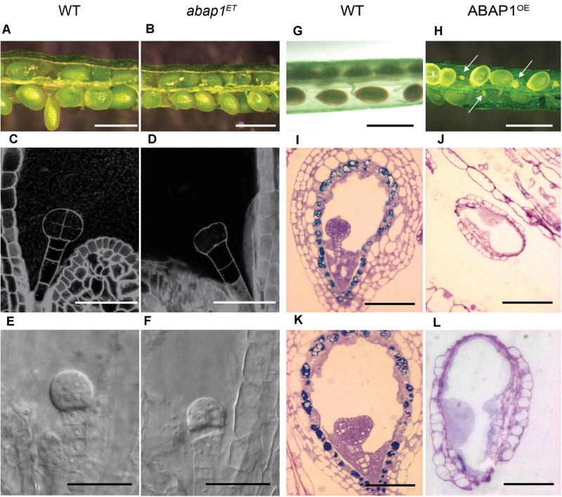
Embryo development in plants with altered levels of ABAP1. Images of siliques at the same developmental stages, comparing embryo and seed development in wild-type (WT) and mutant plants. Left panels (A–F) shows comparison between WT and abap1ET plants. (A,B) Open siliques of WT and abap1ET plants showing normal seed size. (C–F) Initial events in embryo development from WT and abap1ET seeds. Dark field illumination microscopy of WT seeds with embryo at 8 cell stage (C), and abap1ET seeds with the embryo at the initial zygotic divisions (D). Nomarski microscopy of embryos from WT plants at globular cell stage (E) and from abap1ET plants with defects after the initial zygotic divisions (F). Right panels (G–L) shows comparisons between WT and ABAP1OE plants. (G) Open WT siliques with normal developing seeds; and (H) open ABAP1OE siliques with small and shriveled seeds (arrows). (I,K) Micrographs of sections of WT seeds with embryos at globular (I) and transition to early heart stages (K). (J,L) Micrographs of sections of ABAP1OE seeds that did not develop and/or arrested before fertilization (J). (L) is a higher magnification of (J). Scale bars: 1 mm in (A,B,G,H); 20 μm in (C–F); 100 μm in (I–K); and 50 μm in (L). For abap1ET and WT Landsberg erecta background plants, a total of 300 seeds in 15 siliques were analyzed for each genotype. For ABAP1OE a total of 708 seeds were analyzed in 36 siliques, and for Col-0 WT plants 432 seeds were analyzed in 18 siliques.

