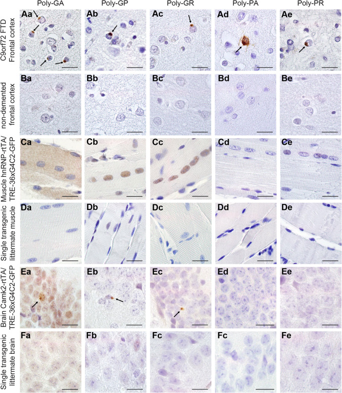
Fig. 2.
Expression of 36× G4C2 human repeats is sufficient to evoke sense DPR formation in vivo. (A,B) Human prefrontal cortex of C9FTD patients (A) or non-demented controls (B) were used as positive and negative controls for the detection of DPR pathology. Arrows indicate perinuclear aggregates of DPRs. (C) In TRE-36×G4C2-GFP/hnRNP-rtTA DT mice, poly-GA showed both diffuse cytoplasmic and nuclear localization, whereas diffuse poly-GP and poly-GR were observed in the nucleus of the EDL muscle. (D,F) ST littermates received the same dox treatment and contained only one transgene, either TRE only or rtTA only, and were all negative for DPRs. (E) TRE-36×G4C2-GFP/Camk2-alpha-rtTA DT mice showed some sparse perinuclear aggregates of sense DPRs in the hippocampus dentate gyrus (indicated by arrows). DPR staining was performed on all mice in this study. ST, 4 weeks dox, n=15; DT, 4 weeks dox, n=16. Scale bars: 20 µm.

