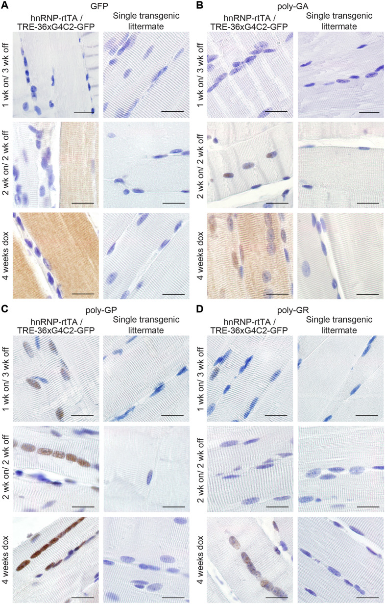
Fig. 5.
GFP and sense DPRs are reduced after dox withdrawal. (A) GFP staining of EDL muscle of TRE-36×G4C2-GFP/hnRNP-rtTA DT mice showed a reduction in the intensity of staining when mice received 1 or 2 weeks of dox water, followed by 2-3 weeks of normal drinking water compared to DT littermates that received 4 weeks of dox. (B) Poly-GA staining of EDL muscle showed clearance of cytoplasmic poly-GA in all washout groups but retention of nuclear poly-GA after 2 weeks of dox withdrawal. (C,D) Poly-GP staining is reduced in the nucleus of EDL muscle (C) and Poly-GR staining is cleared from nuclei of EDL muscles after dox withdrawal (D). ST littermates, consisting of either TRE only or rtTA only, received the same dox treatment and were all negative for GFP and DPRs. All stainings were performed on all mice in this study. Numbers per group were: ST, 1 week dox, n=7; DT, 1 week dox, n=8; ST, 1 week on/3 weeks off, n=4; DT, 1 week on/3 weeks off, n=7; ST, 2 weeks dox, n=6, DT, 2 weeks dox, n=8; ST, 2 weeks on/2 weeks off, n=6; DT, 2 weeks on/2 weeks off, n=5; ST, 4 weeks dox, n=15; DT, 4 weeks dox, n=16. Scale bars: 20 µm.

