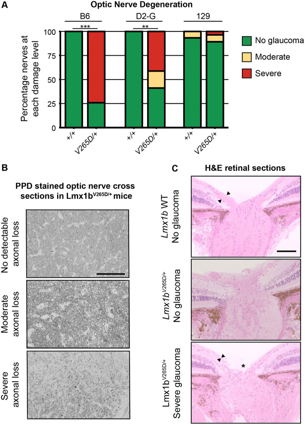
Fig. 5.
B6.Lmx1bV265D/+ mice develop glaucomatous neurodegeneration whereas 129.Lmx1bV265D/+ mice do not. (A) Degree of optic nerve damage evident in PPD-stained cross sections (see Materials and Methods) **P<1.0×10⁻⁵, ***P<1.0×10⁻¹⁰ (Fisher's exact test). (B) Representative images of PPD-stained optic nerve cross sections from Lmx1bV265D/+ mice. Top: healthy nerves at 10 months of age had no detectable axonal damage. These axons had a clear axoplasm and darkly stained myelin sheaths. Middle: moderate optic nerve degeneration with some axon loss and early gliosis. Bottom: severe damage and extreme axon loss with extensive glial scarring. Scale bar: 50 μm (C) H&E-stained optic nerve heads with flanking retina. WT eyes have normal nerve heads with a thick nerve fiber layer (arrowheads), as do unaffected mutants. Severely affected mutants have pronounced optic nerve excavation (asterisk) with loss of the nerve fiber layer (arrowheads). Scale bar: 200 μm.

