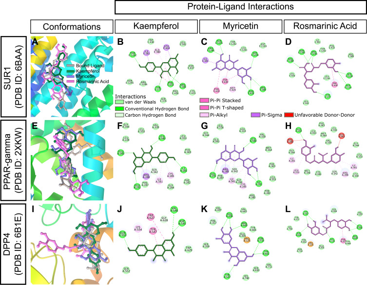
Figure 10.
In silico binding of kaempferol, myricetin and rosmarinic acid to molecular targets of diabetes. (A-D) SUR1, (E-H) PPAR-gamma, and (I-L) DPP4 complexes with ligands. Protein secondary structures are shown as depth-cued cartoons (rainbow) and ligands are shown as sticks. The leftmost columns show best docking poses and other columns depict 2-D representations of protein-ligand interactions.

