Fig. 3.
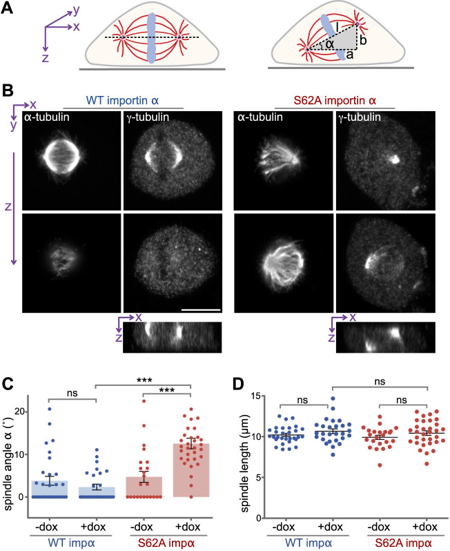
Phosphorylation of importin α controls spindle orientation. (A) Schematic illustration of spindle orientation in tissue culture cells. The axis of a normal spindle is oriented parallel to the coverslip (left), whereas the axis of a misoriented spindle is tilted (right). The angle α of spindle tilt was calculated as: α=arctan (b/a). Spindle length (l) was calculated as the 3D distance between the spindle poles (l2=a2+b2). (B) Importin α S62A causes misorientation of the spindle. NRK cells expressing WT importin α or importin α S62A were arrested at metaphase using MG132 and then dual-stained for α-tubulin and γ-tubulin to label spindle microtubules and the spindle poles, respectively. Z-sections were captured by confocal microscopy. Orthogonal sections through the z-stacks along the x-axis (bottom panels) indicate the relative spindle pole localization on the z-axis. Scale bar: 10 μm. (C) Importin α (impα) S62A results in tilted spindles. The spindle tilt angle α was calculated as shown in A for cells with or without doxycyclin (dox) treatment. n>30. ***P<0.0001; ns, WT −dox versus WT +dox P=0.2478 (Student's t-test). (D) Importin α S62A expression does not alter spindle length. The spindle length l was determined as depicted in A. n>30. ns for WT +dox versus S62A +dox indicates P=0.5676, WT −dox versus WT +dox P=0.185 and S62A −dox versus S62 +dox P=0.196 (Student's t-test). Data are presented as mean±s.e.m.

