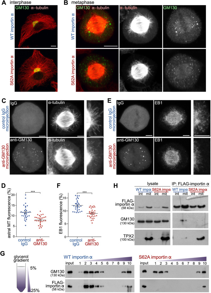
Fig. 6.
GM130 regulates astral microtubule growth via phosphorylated importin α. (A) Importin α S62A does not alter Golgi structure or the microtubule network during interphase. Interphase NRK cells expressing WT importin α or importin α S62A were fixed and stained for α-tubulin and the Golgi membrane protein GM130. Maximum intensity projections are shown. Scale bar: 10 µm. (B) Importin α S62A delocalizes mitotic Golgi clusters from the spindle pole area. NRK cells expressing WT importin α or importin α S62A were arrested at metaphase using MG132, and then fixed and stained for GM130 and α-tubulin. Maximum intensity projections are shown. Scale bar: 10 µm. (C,E) Blocking the GM130-importin α interaction inhibits astral microtubule growth and delocalizes Golgi clusters from the spindle poles. NRK cells were arrested at metaphase for 1 h and microinjected with inhibitory antibodies against GM130 or with control IgG. Injected cells were incubated for another 30 min at 37°C, followed by cold treatment for 5 min. They were then allowed to regrow microtubules for 10 min, before fixation and staining for α-tubulin (C), EB1 (E) and injected IgG. Maximum intensity projections are shown. Enlargements of the astral microtubule area are shown in the panels on the right. Scale bars: 5 µm. (D,F) Quantitation of astral microtubule (MT) fluorescence (D) and astral EB1 fluorescence (F) from the experiments shown in C and E, respectively. Data are presented as mean±s.e.m. n>30. ***P<0.0001 (Student's t-test). (G) Importin α S62A does not associate with mitotic Golgi membranes. Post-chromosomal supernatant of mitotic HeLa cells expressing FLAG-tagged WT importin α or importin α S62A was centrifuged through a linear 5–25% glycerol gradient to separate membrane components by size (left). Ten fractions were collected from the top. The membranes from each fraction were collected by centrifugation and then analyzed by western blotting of the indicated proteins (right). (H) Phosphorylation of importin α shifts its binding preference from TPX2 to GM130. Interphase (int) or mitotic (mit) HeLa cells expressing FLAG-tagged WT importin α or importin α S62A were lysed and incubated with anti-FLAG antibody-conjugated beads (IP, immunoprecipitation). Beads were then washed with lysis buffer and analyzed by western blotting for the indicated proteins. Data in G and H are representative of three experiments.

