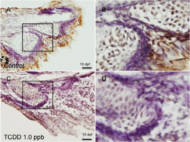
FIG. 7.
PCNA immunoreactivity in medaka hypurals. Five individual embryos were exposed to DMSO or 0.2 ppb TCDD at 4 hpf for 1 h, washed, and incubated in ERM at 26°C. At 10 dpf, embryos were dechorionated, fixed, and processed for sectioning and immunohistochemistry as described in “Materials and Methods.” (A) DMSO-exposed animals; (C) 1 ppb TCDD-treated animals. B and D are magnifications of the rectangular area indicated in A and C, respectively. Black arrows over nuclei indicates PCNA immunoreactivity in component cells of hypurals. White arrows indicate an absence of PCNA immunoreactivity. Fields shown are representative of all individuals in a given group. Bar = 100 μm.

