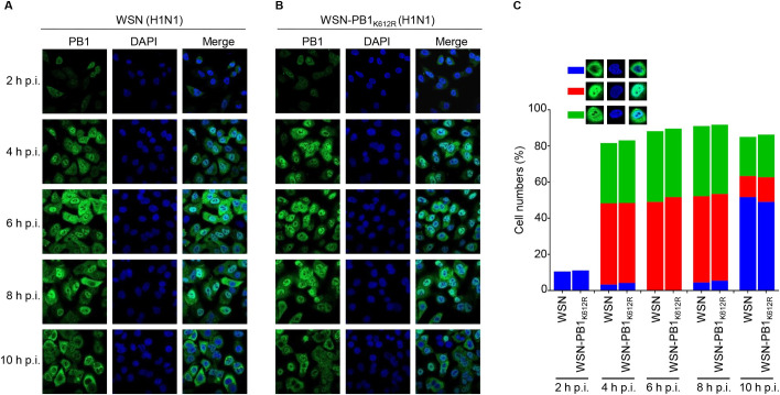
Fig 5. SUMOylation at K612 does not affect the cellular localization of PB1.
(A, B) A549 cells were infected with WSN (H1N1) (A) or WSN-PB1K612R (H1N1) (B) at an MOI of 5, and the cells were fixed at the indicated time points, followed by staining with a rabbit anti-PB1 pAb and incubation with Alexa Fluor 488 donkey anti-rabbit IgG (H+L). The nuclei were stained with DAPI. Representative images are shown. (C) Quantitative analysis of PB1 localization in virus-infected cells. The results shown are calculated from one hundred cells. On the basis of the confocal microscopy in panels A and B, the localization of PB1 was categorized into three types: predominantly cytoplasmic localization (blue), clear nuclear localization (red), and simultaneous localization in the cytoplasm and nucleus (green).

