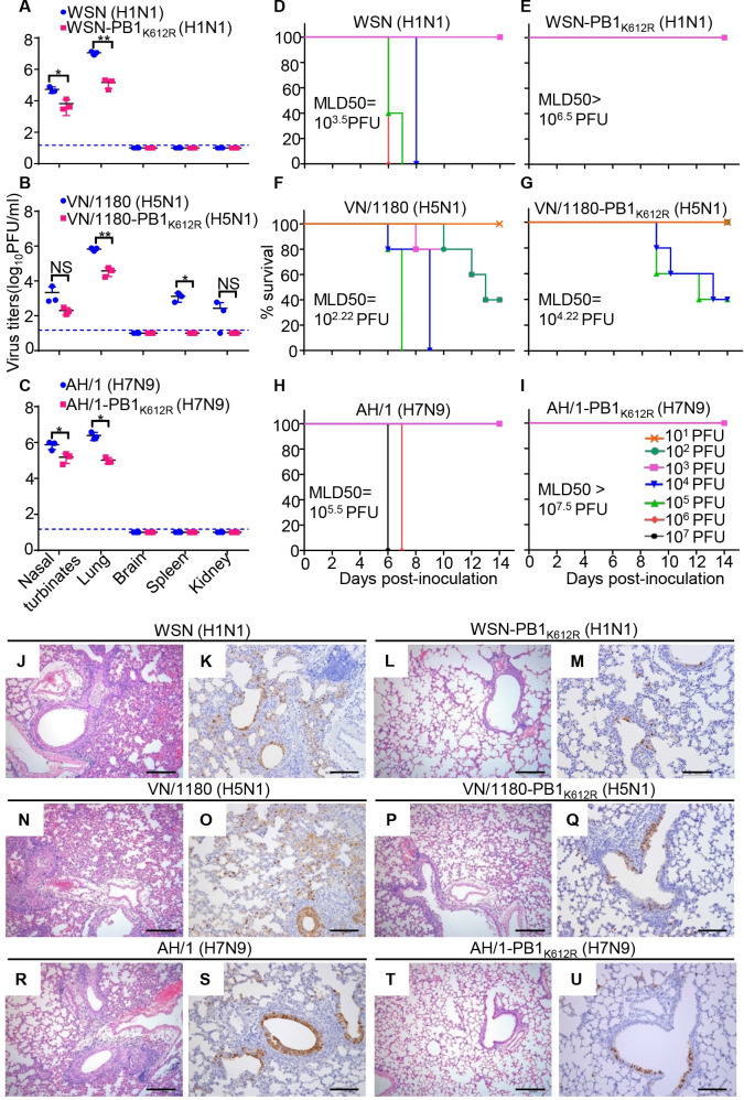
Fig 9. SUMOylation-defective PB1/K612R mutation attenuates the replication and pathogenicity of IAVs in mice.
(A) Replication of WSN (H1N1) and WSN-PB1K612R (H1N1) in mice. Six-week-old BALB/c mice were inoculated intranasally with 106 PFU of WSN (H1N1) or WSN-PB1K612R (H1N1), and organs were collected on day 3 p.i. for virus titration by plaque assays (n = 3). Data shown are mean ± SD for organ samples from three mice; significance was assessed with a one-way ANOVA followed by t-test. *, P < 0.05; **, P < 0.01. NS, not significant. (B) Replication of VN/1180 (H5N1) and VN/1180-PB1K612R (H5N1) in mice inoculated with 105 PFU of the indicated virus, as determined in (A). (C) Replication of AH/1 (H7N9) and AH/1-PB1K612R (H7N9) in mice inoculated with 106 PFU of the indicated virus, as determined in (A). (D, E) Mortality assessment of mice infected with WSN (H1N1) (D) or WSN-PB1K612R (H1N1) (E). Mice were inoculated intranasally with different doses of the indicated viruses and observed for 14 days. (n = 5 mice for each group). (F, G) Mortality assessment of mice infected with VN/1180 (H5N1) (F) and VN/1180-PB1K612R (H5N1) (G) as determined in (D, E). (H, I) Mortality assessment of mice infected with AH/1 (H7N9) (H) and AH/1-PB1K612R (H7N9) (I) as determined in (D, E). (J-M) Hematoxylin-and-eosin (H&E) staining (J, L) and immunohistochemical (IHC) staining for viral NP (K, M) of lung sections prepared on day 3 p.i. from mice intranasally infected with 106 PFU of WSN (H1N1) or WSN-PB1K612R (H1N1). (N-Q) H&E staining (N, P) and IHC staining for viral NP (O, Q) of lung sections prepared on day 3 p.i. from mice intranasally infected with 105 PFU of VN/1180 (H5N1) or VN/1180-PB1K612R (H5N1). (R-U) H&E staining (R, T) and IHC staining for viral NP (S, U) of lung sections prepared on day 3 p.i. from mice intranasally infected with 106 PFU of AH/1 (H7N9) or AH/1-PB1K612R (H7N9). Scale bars = 200 μm (J, L, N, P, R, and T); Scale bars = 100 μm (K, M, O, Q, S, and U).

