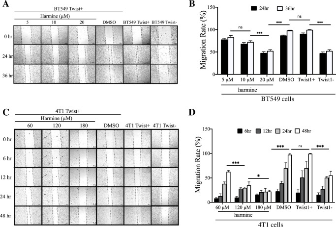
Fig 1. Harmine inhibits migration of breast cancer cells in wound healing assays.
A, BT549 cells were treated with harmine (5, 10, 20 μM) or DMSO (solvent control) and images were captured at different time points (0, 24, and 36 hr). Untreated BT549 Twist+ and BT549 Twist- cells were included as positive and negative controls for migration, respectively. Images are from a single experiment. The scale bars represent 200 μm. B, Quantitative analysis of results from three independent experiments including the one shown in A. Statistical significance was determined based on data from 36 hr. C, 4T1 cells were treated with harmine (60, 120, 180 μM) or DMSO (solvent control) and images were captured at different time points (0, 6, 12, 24, and 48 hr). Untreated 4T1 Twist+ and 4T1 Twist- cells were included as positive and negative controls for migration, respectively. The black particles in images under 120 μM and 180 μM are harmine. Images are from a single experiment. The scale bars represent 200 μm. D, quantitative analysis of results from three independent experiments including the one shown in C. Statistical significance was determined based on the data from 48 hr. Definitions of statistical significance are * p < .05, ** p < .01, *** p < .001, and **** p < .0001.

