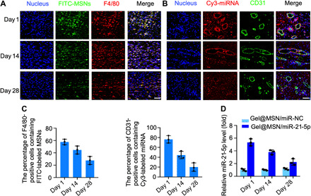
Fig. 7. Time course analysis of the transfection efficiency of Gel@MSN/miR-21-5p in vivo.

MSNs were prelabeled with FITC (green), and miR-21-5p was prelabeled with Cy3 (red). The hydrogel (FITC-labeled Gel@MSN/miR-21-5p or Cy3-labeled Gel@MSN/miR-21-5p) was injected into the mid-myocardium of each target site in the pigs. The duration and efficiency of MSNs and miRNA delivery upon Gel@MSN/miR-21-5p injection were monitored using time course analysis at 1, 14, and 28 days after injection. (A) Histological sections of the infarct region in the Gel@MSN/miR-21-5p group were immunolabeled with the hematoxylin and eosin (H&E) macrophage marker F4/80. (B) Histological sections of the infarct region in the Gel@MSN/miR-21-5p group were immunolabeled with the endothelial marker CD31. Cell nuclei were counterstained with DAPI (blue). (C) F4/80+FITC+ and CD31+Cy3+ double-positive cells were quantified from at least eight high-resolution images acquired from at least eight different regions of each heart. (D) miR-21-5p levels were detected using real-time quantitative PCR at different time points. The transfection efficiency was determined by quantifying the miRNA level. Scale bars, 100 μm. n = 3 per group. The data are shown as means ± SD. Photo credit: Yan Li, Shanghai Ninth People’s Hospital, College of Stomatology, Shanghai Jiao Tong University School of Medicine, Shanghai 200011, China.
