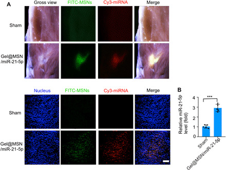
Fig. 8. The MSN/miRNA complex could only be released from Gel@MSN/miR-21-5p at the infarct region.

For examination of on-demand delivery, the hearts were harvested at 28 days after MI for fluorescent imaging, RNA extraction, and real-time quantitative PCR analysis. (A) The fluorescent images showed that there were no transfecting cells detected in the sham group. In contrast, it showed that the area of FITC and Cy3 fluorescence exactly overlapped with the infarct region. (B) Quantification of miR-21-5p levels showed that the MSN/miRNA complex could be highly transfected into cells within the infarct region in vivo. Scale bar, 100 μm. ***P < 0.01. The data are shown as means ± SD. Photo credit: Yan Li, Shanghai Ninth People’s Hospital, College of Stomatology, Shanghai Jiao Tong University School of Medicine, Shanghai 200011, China.
