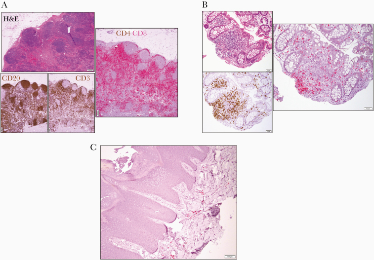
Figure 2.
A, Excisional inguinal lymph node biopsy: hematoxylin and eosin (left upper panel) showing architectural preservation with open sinuses and regressed germinal centers confirmed by immunohistochemical staining for CD20 and CD3 (left lower panels, both in brown) and for combined CD4 (brown) and CD8 (red) staining (scale bar = 200 μm) demonstrating lack of CD4+ cells. B, Cecum biopsy hematoxylin and eosin (left upper panel), showing a lymphoid aggregate in the lamina propria. Immunohistochemical staining for CD3 (left lower panel, brown) and for combined CD4 (brown) and CD8 (red) staining revealing lack of CD4+ cells (scale bar = 50 μm). C, Immunohistochemical staining of lymphocytes in the superficial dermis of a left tibial verrucous plaque for CD4 (brown) and CD8 (red) (scale bar = 100 μm). Abbreviation: H&E, hematoxylin and eosin.

