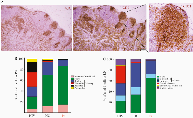
Figure 5.
A, Immunohistochemical staining of an excisional inguinal lymph node biopsy: B cells are highlighted by the CD21 staining (left upper panel), while IgD staining highlights naive or unswitched memory B cells. Insert on right panel highlights the intricate CD21+ mesh of follicular dendritic cells within B-cell primary follicles (scale bar = 50 μm). B, Distribution of B-cell subsets in peripheral blood of HIV-1–infected individuals in the absence of antiretroviral treatment (HIV, average of 3 female donors, median CD4+ T-cell count 551 cells/μL, 27% of total T cells), healthy controls (average of 7 female donors, median CD4+ T-cell count 856 cells/μL, 55% of total T cells), and the proband. The proportion of immature/transitional B cells (CD10+CD27–) is in pink; in green is the proportion of naive B cells (CD21hiCD27–); in dark blue are resting memory B cells (CD21hiCD27+); in red are tissue-like memory B cells (CD21loCD27–); in black are activated memory B cells (CD21loCD27+); and in yellow is the proportion of plasmablasts (CD20–/CD21loCD27++). C, Distribution of B-cell subsets in lymph node of HIV-1–infected individuals in the absence of antiretroviral treatment (HIV, average of 3 female donors, median CD4+ T-cell count 551 cells/μL, 27% of total T cells), healthy controls (average of 7 female donors, median CD4+ T-cell count 856 cells/μL, 55% of total T cells), and the proband. The proportion of naive B cells (IgD+CD38–CD27–) is indicated in green, the proportion of unswitched memory B cells (IgD+CD38–CD27+) in light blue, switched memory B cells (IgD–CD38–CD27+) in dark blue, germinal center B cells (IgD–CD38+) in red, undefined or indeterminate B cells in purple, and plasmablast/plasma cells (IgD–CD38++) in yellow. Abbreviations: HC, healthy control; HIV, human immunodeficiency virus; LN, lymph node; PB, peripheral blood; Pt, proband.

