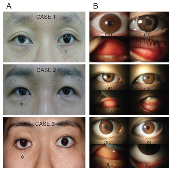
Fig. 3.
Photographs of three patients’ normal eyes and anophthalmic eyes. (A) Gross photos of the three patients at primary gaze. (B) Slit-lamp photos of the normal eye, the ocular prosthesis and the upper and lower conjunctiva of the anophthalmic socket. The ocular prosthesis is marked by an asterisk. OD = right eye; OS = left eye; UL = upper lid; LL = lower lid. Informed consent for publication of the clinical images was obtained from the patients.

