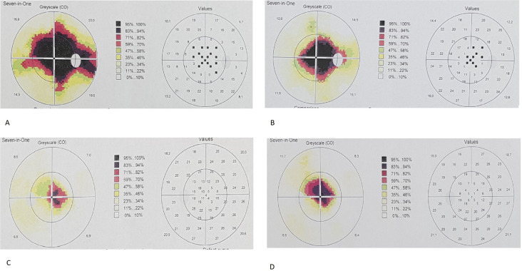
Fig. 2.
Perimetry results of an SMD subject before treatment (A) and one year after treatment (B). Note that central visual field defect decreased during the first year period. Perimetry results of the fellow eye of the same subject during the study period (C, D). Note the worsening of the central visual field defect.

