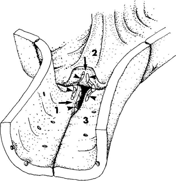
Figure 1.

Sketch of an endothelial tricellular junction in a pulmonary capillary, based on freeze fracture electron micrographs. The flap of cell “2” has been peeled back and the gap between cells “1” and “3” has been widened at its end in order to show the tight junction strands (arrowheads). Those on the underside of the flap join with those on the luminal surface of cells “1” and “3”. Those on the edge of cell “1” join with corresponding structures on the edge of cell “3” (which are not shown). Even with the flap lowered and the tight junctions properly formed, a patent channel remains (arrows).
Reproduced from Fig. 13 of Walker et al. 1994,45 Copyright, with permission from Elsevier.
