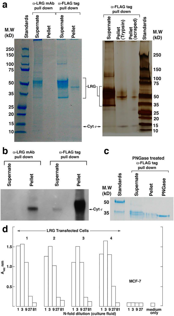
Figure 4. Lrg1-transfected MCF-7 cells express variably glycosylated forms of LRG1 most of which are secreted, while a 45 kD form capable of binding Cyt c is retained in the cells.
(a) A heterogeneous population of LRG1 polypeptides visualized by SDS-PAGE was immunoprecipitated from the supernate of lrg1-transfected cells employing antibody-coupled beads specific for LRG (2F5.A2) and the FLAG tag present in recombinant LRG1. On the left the gel was stained with GelCode Blue and on the right a separate gel was silver stained. Major bands were identified as LRG1 by MS (see Table I). A uniquely-sized polypeptide (45 kD) was retained in the cell pellet. Co-immunoprecipitation of Cyt c was also apparent.
(b) Western blotting confirmed the presence of Cyt c in the immunoprecipitates in “a.”
(c) PNGase F reduced the molecular weight of LRG1 present in both the supernate and cell pellet to a size approximating the non-glycosylated polypeptide (34 kD).
(d) Indirect ELISA demonstrated the presence of LRG1 in the supernates of four clones of lrg1-transfected MCF-7 cells but not in the supernate of non-transfected cells.

