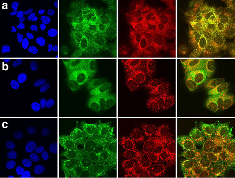
Figure 5. Intracellular localization of LRG1 in parental MCF-7 cells and in cells transfected with lrg1 by confocal immunofluorescent microscopy.
(a) In MCF-7 cells, LRG1 (green) co-localized with the Golgi marker, GRASP 65 (red). LRG was detected with LRG1 peptide-specific mAb 3C9.D5. Nuclei (blue) were stained with DAPI (left panel). In the far right panel the green and red fluorescent images are merged.
(b) In lrg1-transfected MCF-7 cells, LRG1 detected with mAb 3C9.D5 (green) co-localized with GRASP 65 (red) and also appeared in a diffuse pattern outside the Golgi.
(c) Recombinant LRG1 detected with an anti-FLAG tag mAb (green) partially co-localized with GRASP 65 (red) in lrg1-transfected MCF-7 cells and was also prominent just below the cell surface consistent with its secretion from lrg1-transfected cells (see Fig. 4d).

