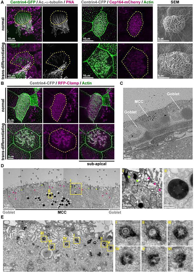
Fig. 5. Cellular changes in trans-differentiating MCCs.
(A) Confocal and scanning electron microscopy (SEM) micrographs of normal and trans-differentiating MCCs reveal disorganized basal bodies (Centrin4-GFP, green / Centrin4-CFP, grey), cilia de-acetylation (Ac.-α-Tubulin, grey), F-actin remodeling (Actin, green) and loss of basal body distal appendages (Cep164-mCherry) during MCC cilia retraction. Cf. Fig. S5E for quantifications. Centrin4-GFP/Ac.-α-Tubulin/PNA N = 1 embryo; N = 8 normal MCCs; N = 2 trans-differentiating MCCs. Centrin4-CFP/Cep164-mCherry/Actin N = 3 embryos; N = 15 normal MCCs; N = 15 trans-differentiating MCCs. SEM N = 2 embryos; N = 15 normal MCCs; N = 11 trans-differentiating MCCs. (B) Confocal micrographs of a normal and a trans-differentiating MCC stained for basal bodies (Centrin4-CFP, gray), rootlets (RFP-Clamp, magenta) and F-actin (Actin, green) reveals loss of basal body alignment as well as apical and sub-apical F-actin organization. N = 8 embryos. (C,D) Transversal sectioning and transmission electron microscopy (TEM) shows normal and abnormal basal bodies (green arrowheads), mucus granules (magenta arrowheads) and an enrichment of lysosomes (white arrowheads) in a trans-differentiating MCC. Magnified areas are indicated by yellow boxes. Image in D was reconstructed from multiple individual micrographs of cell depicted in C. N = 1 embryo. (E) Parallel section and TEM shows normal and abnormal basal bodies in a trans-differentiating MCC. Magnified areas are indicated by yellow boxes. N = 1 embryo.

