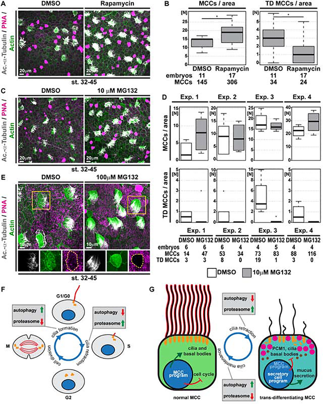
Figure 6. Activation of autophagy or inhibition of proteasomal degradation prevents cilia retraction and MCC loss.
(A,C,E) Confocal micrographs of the epidermis stained for MCCs (Ac.-α-Tubulin, grey), F-actin (Actin, green) and mucus (PNA staining, magenta) demonstrate an increase in MCCs and reduced cilia loss in Rapamycin (A) and MG132 (C,E) treated tadpoles. (A) DMSO N = 11; Rapamycin N = 17 embryos. (C) DMSO N = 20; MG132 N = 19 embryos. (E) Altered F-actin and cell morphology was observed even in fully ciliated MCCs after MG132 treatment. DMSO N = 20; MG132 N = 5 embryos. (B,D) Quantification of total MCC numbers and number of MCCs showing trans-differentiation (TD) morphology. Mann Whitney test, * P < 0.05. Due to greater variability between individual experiments with 10μM MG132, individual replicates are depicted separately in (D) for transparency. (F,G) Schematic summary of protein degradation pathways in primary cilia (E) and MCC cilia (F) maintenance and retraction.

