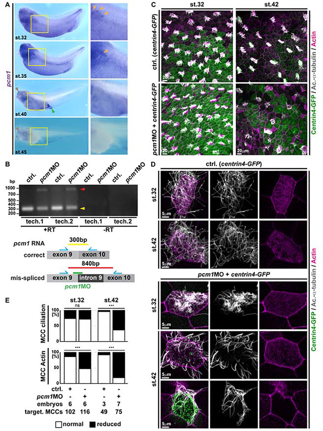
Figure 7. Loss of PCM1 leads to premature MCC de-ciliation during trans-differentiation.
(A) In situ hybridization shows pcm1 (purple) expression in the epidermis and other multiciliated areas (nephrostomes, orange arrowheads; nasal pit, brown arrowheads; cloaca, green arrowhead) and progressive loss of expression in the epidermis during stages (32 to 45) of MCC trans-differentiation. Yellow boxes indicate location of magnified images. St. 32 N = 13; st. 35 N = 7; st. 40 N = 7; st. 45 N = 12 embryos. (B) RT-PCR in controls and pcm1MO injected specimens confirms pcm1 intron 9 retention after injection of the splice-blocking MO. Schematic representation of correct and mis-spliced transcripts, expected amplicons and localization of primers (blue) as well as MO-targeting (green). N = 3 biological and 2 technical replicates each. (C-D) Confocal micrographs of centrin4-GFP injected (basal bodies, green) control (ctrl.) and pcm1MO treated MCCs reveal normal ciliation (Ac.-α-Tubulin, grey) and only mild effects on apical F-actin organization (Actin, magenta) at stage 32, but reduced ciliation and severe apical F-actin defects in targeted MCCs already shortly after onset of trans-differentiation at stage 42. Ctrl. st. 32 N = 6; ctrl. st. 42 N = 3; pcm1MO st. 32 N = 6; pcm1MO st. 42 N = 7 embryos. (E) Quantification of ciliation and F-actin defects in targeted MCCs at stages 32 and 42. χ2 test, ns P > 0.05 = not significant, *** P < 0.001.

