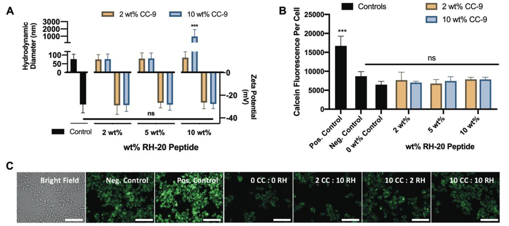
Figure 6: Dual peptide-modified nanogels fail to disrupt endosomes.
(A) Nanogel modification with both CC-9 and RH-20 to different extents (2 – 10 wt%) did not significantly influence the nanogel zeta potential or hydrodynamic diameter, and led to aggregation only in the case of the 20 wt% (equal weight CC-9 and RH-20) conjugate (control = unmodified nanogels, z-average diameter ± PDI width, zeta potential ± zeta deviation, n = 3). (B) Dual peptide-modified nanogels fail to facilitate endosomal escape in SW-48 cells, suggesting that enhanced nanogel co-localization with 10 wt% CC-9 is insufficient to enable endosome disruption by RH-20 (positive control = 200 μg / mL RH-20, negative control = calcein and media only, 0 wt% control = unmodified nanogels in calcein solution). (C) Representative images for SW-48 cells in each incubation condition, labeled by peptide modification cocktail (green = calcein, scale bar = 100 μm). Images are enlarged and reprinted in Figure S4 (mean fluorescence ± sd, n = 3, ***p < 0.001, 2-way ANOVA).

