
Fig. 2.
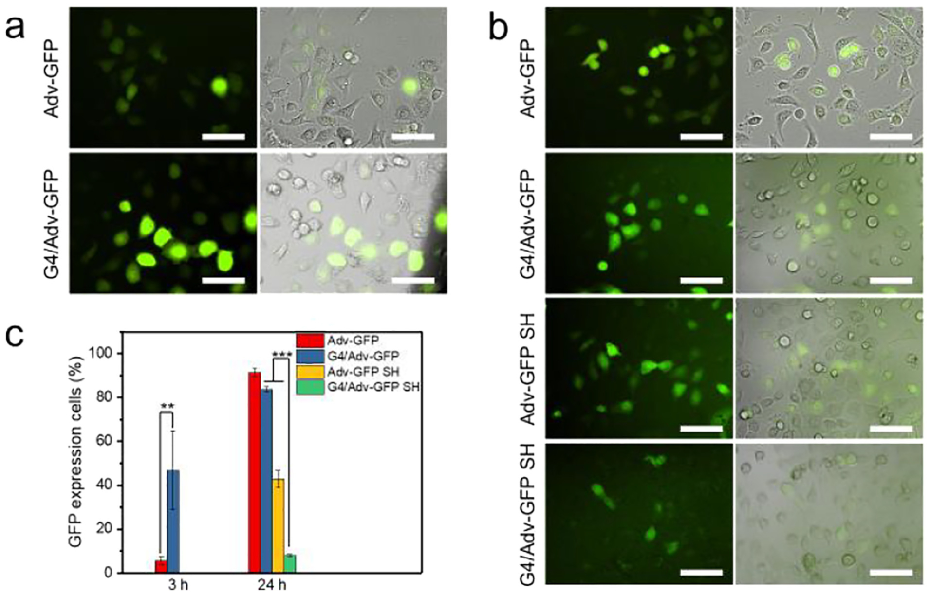
In vitro transfection efficiency of Adv-GFP. a) Fluorescence images of cells incubated with Adv-GFP or G4/Adv-GFP for 3 h, scale bar = 100 μm. b) Fluorescence images of cells incubated with Adv-GFP, G4/Adv-GFP, Adv-GFP SH, or G4/Adv-GFP SH for 24 h, scale bar = 100 μm. c) Quantitative analysis of GFP-expressing cells incubated with Adv-GFP, G4/Adv-GFP, Adv-GFP SH, or G4/Adv-GFP SH by FACS. **, p <0.01. ***, p <0.001.