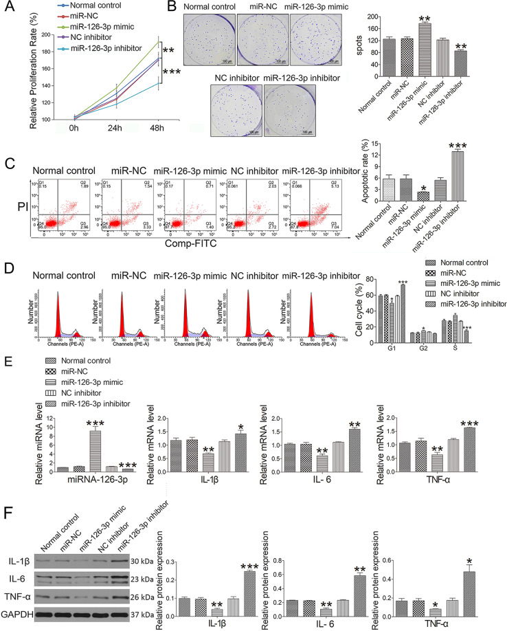
Fig. 2. Effect of miR-126-3p administration on chondrocytes.
A Cell proliferation was determined by CCK-8 at concentrations of 100 pM (miRNA-126-3p mimic and inhibitor) for 24 and 48 h. B The colony formation assay was used to analyze the effect of miRNA-126-3p mimic (100 pM) and miRNA-126-3p inhibitor (100 pM) on the proliferation of chondrocytes. C Apoptotic index was determined using flow cytometry in the presence of miR-126-3p mimic (100 pM) or inhibitor (100 pM) constructs for 24 h. D Effect of miR-126-3p on cell cycle progression in the presence of miR-126-3p mimic (100 pM) or inhibitor (100 pM) constructs for 24 h. E The miRNA (IL-1β, IL-6, and TNF-α) and mRNA (miRNA-126-3p) expression was detected by qRT-PCR in the presence of miR-126-3p mimic (100 pM) or inhibitor (100 pM) constructs for 24 h. F Western blot assay was used to detect inflammation-related proteins (IL-1β, IL-6, and TNF-α) in the presence of miR-126-3p mimic (100 pM) or inhibitor (100 pM) constructs for 24 h. Data were expressed as mean ± SEM (n = 3). *P < 0.05, **P < 0.01, and ***P < 0.001 vs. normal control.

