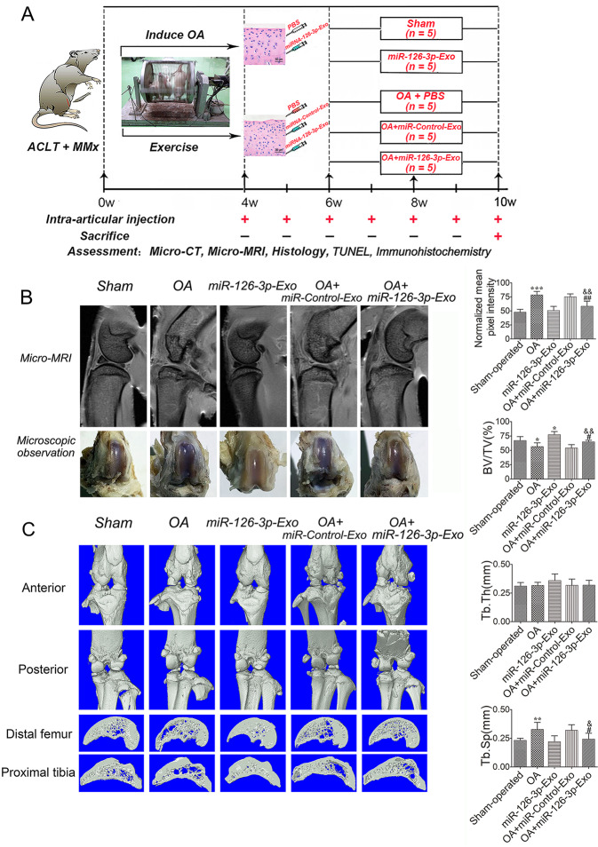
Fig. 4. An overview of study timelines and the rescue of cartilage degeneration by SFC-miR-126-3p-Exos in OA model rats.
A ACLT + MMx rats were put into an electronic rotator cage for 30 min per day as a means of inducing the OA model beginning 1-week post-surgery. At 4 weeks post-surgery, animals were intra-articularly injected with 40 μl of 500 μg/ml SFC-miR-126-3p-Exos. PBS was used as a control in sham and OA-model animals. At 10 weeks post-surgery, the micro-MRI, micro-CT, histology, TUNEL assay, and immunohistochemistry were used as evaluation criteria. B Pixel intensity of a sampled region of epiphyseal trabecular bone and gross morphological were assessed by micro-MRI to quantify the presence of bone marrow lesions in sham operation, SFC-miRNA-126-3p-Exos, OA-induction, OA + SFC-control-Exos, and OA + SFC-miRNA-126-3p-Exos, groups. C Representative micro-CT three-dimensional reconstructions of tibial and femoral subchondral bones in the rats after ACLT + MMx surgery. Micro-CT images of animals in each group were used for measurements of BV/TV, Tb. Th and Tb. Sp. Data were expressed as mean ± SEM (n = 5). *P < 0.05, **P < 0.01, and ***P < 0.001 vs. sham-operated group; #P < 0.05 and ##P < 0.01 vs. OA induction group; &P < 0.05 and &&P < 0.01 vs. OA + miR-Control-Exos group.

