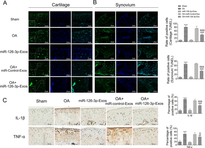
Fig. 6. Assessment of apoptosis and inflammation in cartilage mediated by SFC-miR-126-3p-Exos in OA model rats.
A At 10 weeks post-surgery, animals were sacrificed and the knee joints were collected for TUNEL analysis. A representative TUNEL-stained section of articular cartilage was used to assess apoptosis. B Representative TUNEL-stained section of synovial tissues was used to assess apoptosis. C The immunohistochemical stainings of IL-1β and TNF-α were performed in OA model rats. The ratios of immunoreactive positive cells were analyzed. Data were expressed as mean ± SEM (n = 5). Data were expressed as mean ± SEM (n = 5). ***P < 0.001 vs. sham-operated group; ##P < 0.01 and ###P < 0.001 vs. OA induction group; &P < 0.05, &&P < 0.01, and &&&P < 0.001 vs. OA + miR-Control-Exos group.

