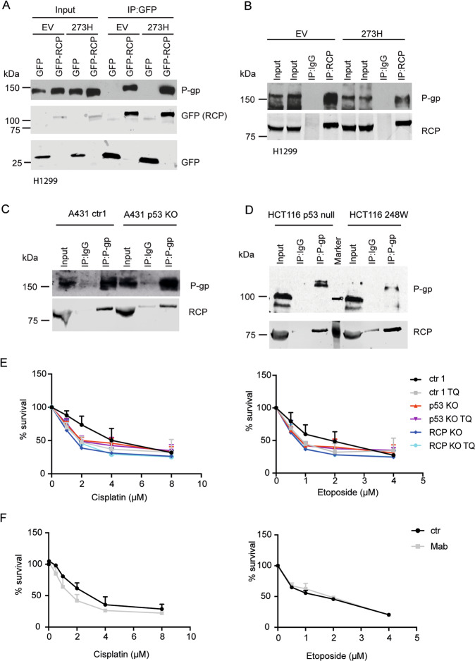
Fig. 3. RCP interacts with P-gp.
A H1299 cells, stably expressing mutant p53 R273H or an empty vector (EV) (PCB6) were transfected with GFP or GFP-RCP and cell lysates were immunoprecipitated with GFP. P-gp and GFP expression was analysed by western blot. B Cell lysates of H1299 cells stably expressing mutant p53 R273H or an EV (PCB6) were immunoprecipitated with RCP or an IgG control. P-gp and RCP expression was analysed by western blot. C P-gp and IgG were immunoprecipitated from A431 ctr or A431 KO cells. Endogenous P-gp and RCP protein levels were analysed by western blot. D P-gp and IgG were immunoprecipitated from HCT116 -/- or 248W cells. Endogenous P-gp and RCP protein levels were analysed by western blot. E Resazurin survival assay of ctr, p53 KO or RCP KO cells treated with increasing doses of cisplatin and etoposide in combination with 0.5 µM Tariquidar or DMSO control for 96 h. Statistical differences were measured using a paired two-way ANOVA corrected for multiple testing (two-sided) and IC50 values were measured using a linear regression analysis. IC50 and P-values can be found in Supplemental Tables 6 and 7. Error bars indicate SD of average values of three independent experiments. F Resazurin survival assay of ctr1 A431 cells treated with increasing doses of cisplatin or etoposide in the presence or absence of Mab16 (1 µg/mL) for 96 h. Statistical differences were measured using a paired two-way ANOVA (two-sided) corrected for multiple testing and IC50 values were measured using a linear regression analysis. IC50 and P-values can be found in Supplemental Table 8. Error bars indicate SD of average values of three independent experiments.

