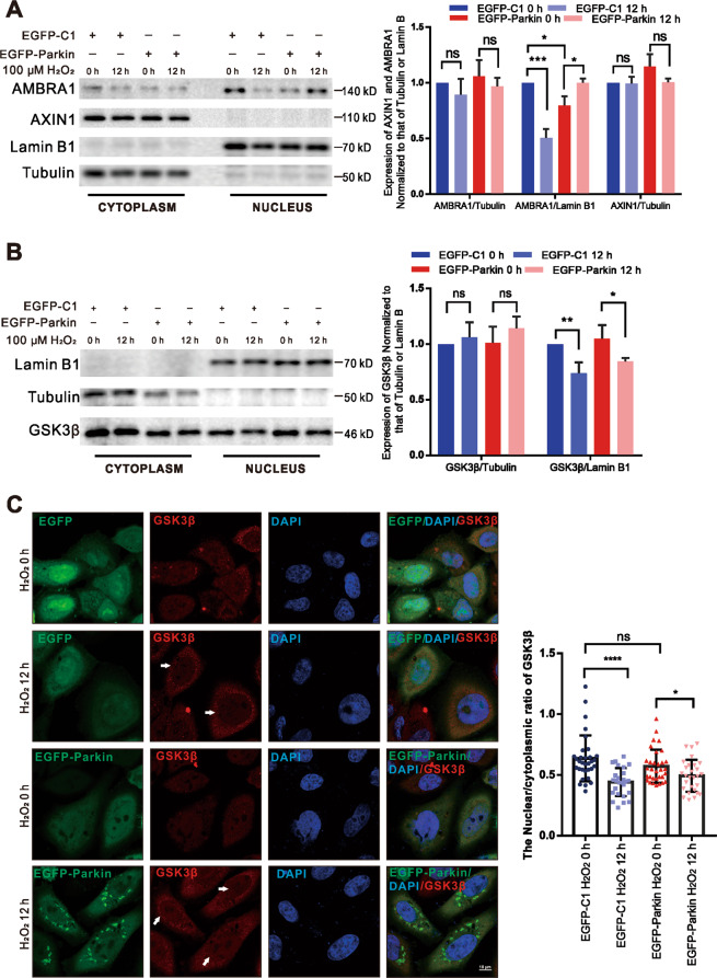
Fig. 5. H2O2 stimulation promotes the expression of c-Myc by decreasing GSK3β levels in the nucleus.
A, B Cells were transfected with EGFP-C1 or EGFP-Parkin and then stimulated with 100 μΜ H2O2 for 0 h or 12 h. Then, nuclear and cytoplasmic proteins were isolated for WB analysis. The protein levels were analyzed with the indicated antibodies. N = 3; ns, not significant; *P < 0.05; **P < 0.01; ***P < 0.001. The data are from three independent tests and are presented as the mean ± SD. C Cells transfected with EGFP-C1 (green) or EGFP-Parkin (green) were incubated with 100 μΜ H2O2 for 0 h or 12 h. The cells were immunostained with an anti-GSK3β antibody (red). The white arrows show GSK3β in the nucleus. The fluorescence intensity was analyzed with ImageJ, and the data were subjected to statistical analysis (~30 cells for each analysis). Scale bars, 10 μm. Ns, not significant; *P < 0.05; ****P < 0.0001.

