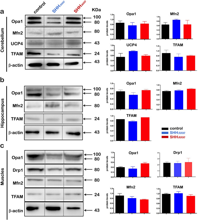
Figure 7.
Protein expression levels showed a trend similar to those of the mRNA levels. (a) Normal mice were sacrificed after SHH5000 or SHH8000 treatment for 16 h. Each cerebellum was collected and immediately stored in liquid nitrogen. Western blotting was then performed to detect differences in mitochondrial-related protein expression levels (Mfn2, Opa1, TFAM, UCP4). (b) Western blotting was performed to detect differences in mitochondrial-related protein expression levels (Mfn2, Opa1, TFAM) in each mouse hippocampus after SHH5000 or SHH8000 treatment. (c) Western blotting was performed to detect differences in mitochondrial-related protein expression levels (Mfn2, Opa1, TFAM) in the muscles of mice after SHH5000 or SHH8000 treatment. Data are analyzed by Image J software, and are presented as the mean ± SD. *P < 0.05, **P < 0.01, ***P < 0.001, compared with the control group, n = 5 per group. Full-length blots/gels are presented in Supplementary Fig. 1a–c. Mfn2, mitochondrial fusion protein 2, a mitochondrial fusion factor on the mitochondrial outer membrane; Opa1, Optic atrophy1, a mitochondrial fusion factor on the mitochondrial inner membrane; TFAM, mitochondrial transcription factor; UCP4, uncoupling protein 4, a mitochondrial stress factor; SHH5000,simulated hypobaric hypoxia at an altitude of 5000 m; SHH8000, simulated hypobaric hypoxia at an altitude of 8000 m.

