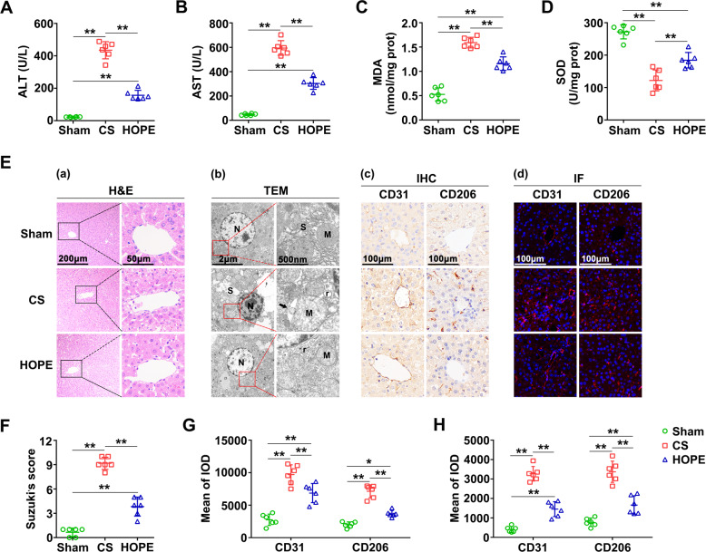
Fig. 1. HOPE improves liver function and alleviates DCD liver injury in rats.
A, B ALT and AST activities in the perfusate. Data are mean ± SD, **P < 0.01 by one-way ANOVA followed by Tukey’s test. n = 6 per group. C, D MDA and SOD activities in rat liver tissues. Data are mean ± SD, **P < 0.01 by one-way ANOVA followed by Tukey’s test. n = 6 per group. E (a) Histological damage in liver of rats. H&E staining was performed on paraffin-embedded section of rat liver tissues. Scale bar = 200 μm (left panels) and 50 μm (right panels). E (b) Changes in the microstructure of hepatocytes. The changes of hepatocellular microstructure were observed using TEM. N, nucleus; M, mitochondria; r, rough endoplasmic reticulum; S, smooth endoplasmic reticulum. The black arrows indicate the lacuna between the inner and outer membranes of mitochondria. Scale bar = 2 μm (left panels) and 500 nm (right panels). E (c, d) Activation of LSECs. IHC staining (c) and IF staining (d) of CD31 and CD206 were performed on paraffin-embedded section of rat liver tissues. Scale bar = 100 μm. F Histological scores were analyzed based on Suzuki’s criteria. Data are mean ± SD, **P < 0.01 by one-way ANOVA followed by Tukey’s test. n = 6 per group. G, H IHC and IF stained fluorescence of CD31 and CD206 were quantified using Image-pro plus 6.0. Data are mean ± SD, *P < 0.05 and **P < 0.01 by two-way ANOVA followed by Tukey’s test. n = 6 per group.

