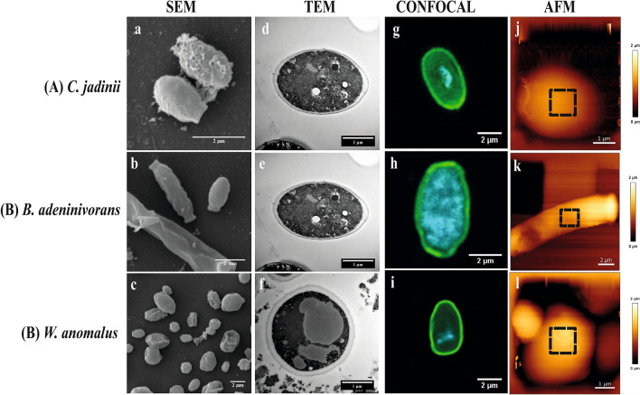
Figure 3.
Cell surface architecture of three inactivated yeast species grown on sugars from lignocellulosic biomass. The pictures show Scanning Electron Microscopy (SEM; a–c), Transmission Electron Microscopy (TEM; d–f), Confocal microscopy (stained with concanavalin A-FITC for mannan) (g–i) and Atomic Force Microscopy (AFM; height) (j–l) micrographs of Cyberlindnera jadinii (panel A), Blastobotrys adeninivorans (panel B) and Wickerhamomyces anomalus (panel C). The SEM and TEM micrographs were taken on yeast creams (before drying), whereas the confocal and AFM micrographs were taken on dried yeast samples, as described in the ‘Material and Methods’. The dotted squares on the AFM height micrographs represent the spots where mapping was done for determination of the Young modulus and measurement of adhesion events, as described in ‘Material and Methods’.

